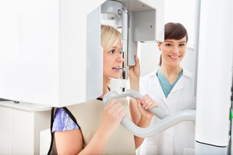
Cone Beam CT Technology
Dental Services
X-rays have long been replaced by CT (computed tomography) scans in dentistry and in other medical fields. Once upon a time, a CT scanner was the latest imaging device that impressed medical practitioners. But now there’s the new cone beam CT technology, although it’s not technically brand new. It’s been used in Europe since 1999 and launched in the US in 2001. It just seems new because this technology is now more commonly used in dental offices.
Lower Radiation Exposure
The new cone beam CT technology was named as such because it uses an x-ray beam that’s shaped like a cone. In contrast, standard CT scanners use a linear fan beam. This improvement means that taking multi-planner views needs only a single revolution around the patient. This minimizes the exposure time, and radiation levels as the entire process require only 3.6 to 6 seconds.
High Accuracy
Dentists prefer CBCT scans because they also offer 3D images from every angle and in real-time. The scans are accurate, come in high resolution, and contain a great amount of detail. A typical CBCT scanner is also compact, while the patient can sit down while the scans are being generated.
This CBCT is perfect for taking images of the craniofacial structures because they offer an extremely high accuracy rate. The best conventional medical CT scanner is accurate to within 0.5 mm, and more typical CT scanners can have a larger margin of error. But for CBCT scanners, the accuracy is within 0.1 mm. That’s at least 5 times more accurate. It’s this level of accuracy that more dentists are relying on to diagnose patients, plan treatment solutions, and educate patients.
Uses in Dentistry
Due to the maxillofacial imaging capabilities and advantages offered by CBCT scanners, they’re ideal for a long list of dental care applications. Both GP practitioners and specialists can use them to assess facial bones and jaws for pathology. They can check for impactions and fractures. They can use these scans to find tumors along with congenital and developmental deformities.
CBCT capabilities can be used in a multitude of ways in dentistry:
Orthodontics
CBCT imaging allows for more accurate planning of treatment and more comprehensive diagnoses. It enables:
- 3D assessments of impacted tooth anatomy and position
- 3D views of crucial structures
- True 1:1 imaging for orthognathic (corrective jaw) surgery treatment planning and growth assessments
- Planning for how dental implants can be placed to replace missing teeth or to function as anchors, and for the placement of temporary anchors.
- Assessing airways.
Implantology
With such accurate 3D scans, dental specialists can make the best plans for the placements of dental implants. These scans are useful throughout the whole process, starting from the diagnosis and treatment to post-operative examinations. It lets dentists do the following:
- Choose the most ideal implant type and size
- Shorten the surgery time
- Boost the confidence of the patient
- Check the distance to other anatomical structures.
- Measure and visualize alveolar bone width and contours
- See if a sinus lift or a bone graft is required.
Basically, the dentist can regard each dental implant case with the confidence borne of knowing that the best imaging equipment has been used for the task.
Studying Sinuses and Airways
The CBCT technology can compile a large amount of data so that the dentist can visualize the sinuses, along with the whole airway path from the nose and the mouth up to the laryngeal spaces. The data can then be used for:
- Finding out about the presence of polyps and the degree of infection
- Determining the borders of anatomic structures
- Calculating the real airway space volume
- Finding the exact location of the airway constriction
- Helping in the diagnosis and treatment of obstructive sleep apnea.
Visualizing for Pathology
Dentists are now better able to study and visualize the pathological processes in the mandible and maxilla, using CBCT scanners. It’s crucial for making surgical plans for resection or biopsy. The info can be used to:
- Provide 3D images of hard tissue abnormalities
- Offer more precise data regarding the location and size of adjacent anatomic structures
- Check the continuing progression of the pathology
- Use multiple scans to monitor the success rate of the treatment.
Impactions
Impacted teeth come with risks, but these risks can be minimized by the more precise 3D rendering of CBCT scanners. The dentist can:
- Use the more accurate 3D analysis to determine if it’s better to treat or not to treat the impacted tooth
- Gather more accurate info regarding the position of the impacted tooth, in relation to the adjacent teeth, their roots, and other crucial anatomic structures.
TMJ
It’s not easy to evaluate the temporomandibular joint (TMJ) with standard radiographs because the images of other structures can be superimposed in the scan images. But it is better with the Cone Beam CT scanner, as the dentist can:
- Evaluate the TMJ condylar anatomy without any image distortion or the superimposition of other images
- Form a more accurate evaluation by getting an accurate 1:1 image ratio.
Endodontics
Admittedly, it’s more practical and more effective to use the standard radiography for common endodontic procedures. However, CBCT scans can offer many types of views (sagittal, coronal, serial axial) which standard radiography can’t provide for dentists. With CBCT scans, the dental specialist can also minimize or even eliminate the superimposition of other non-relevant structures. This lets the dentist get a cleared 3D image of the relevant areas.
The potential endodontic benefits using CBCT include:
- Getting a clear image of typically less accessible root canal systems and hidden internal pulpal anatomy
- Being able to more precisely identify and diagnose periapical endodontic pathosis
- Evaluating the external and internal root resorptive processes
- Determining the root fractures and the other spots of oral trauma
- Better planning before surgeries
- More accurate and comprehensive comparisons of the periradicular bone after the endodontic treatment to see whether the treatment as a failure or a success.
Oral Surgery
Oral surgeons can use CBCT not just for placing dental implants but for making diagnoses and coming up with treatment plans. The oral surgeon can:
- Plan maxillofacial surgeries by getting 3D computer images of the soft and hard tissues
- Find the exact 3D position of the tooth within the alveolar bone, and see how this particular position relates to crucial structures for impactions and extractions.
- Monitor the changes in the airways, skeletal structures, and in the healing reactions
- Produce actual-size CAD-CAM computer images to plan and prepare for surgeries.
Periodontics
Dentists who do periodontal surgery often encounter surprises during the procedure because standard 2D x-rays aren’t as informative and accurate. But with CBCT, the dentist can:
- Evaluate the extent of all furcation movements
- Check all side of all the teeth for periodontal bone defects
- Monitor the rate of periodontal bone loss
- Plan for the use of dental implants by evaluating various bone factors like bone density and size.
- Visualize crucial anatomic structures before the implant or periodontal surgery.
Clearly, the use of cone beam CT technology is becoming more popular for good reasons. Dental specialists need accurate information and more precise imagery for a wide range of dental tasks, and you can achieve this using CBCT technology.
Dr. Farman, the president of the American Academy of Oral and Maxillofacial Radiology, has high hopes for the increasing popularity of cone beam computed tomography scanners. He appreciates the availability of high-resolution scans that require very low levels of radiation. They’re also ideal for use with younger patients, as numerous imaging scanners are mostly designed to serve only the need of adult patients. Dr. Farman says that these scanners can be of great help for both general dentistry and specialized dental care.


